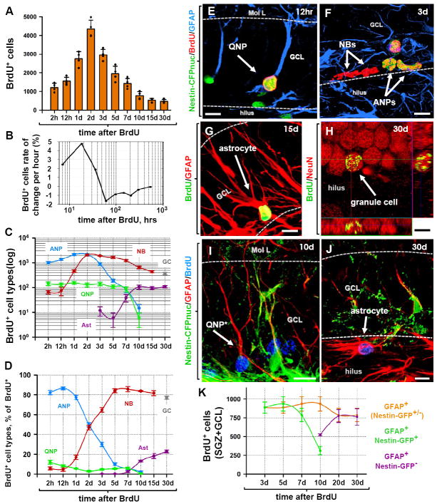
Fig. 4. The fate of dividing cells in the DG.
(A) Time course of changes in the number of BrdU-positive cells after pulse labeling. 2 month old Nestin-CFPnuc mice (n=4 per time point) received a single injection of BrdU (150 mg/kg) and the number of BrdU-labeled cells in the DG was monitored over the course of 30 days.
(B) Rate of changes in the number of BrdU+ cells, reflecting periods of active proliferation, rapid loss, and slower loss of the newborn cells.
(C, D) Time course of changes in defined classes of progenitors and mature cells in the DG of Nestin-CFPnuc mice. BrdU+ cells were immunophenotyped to determine the numbers of labeled cells in defined classes. The results for QNPs, ANPs, NBs, mature neurons (granule cells, GC), and stellar astrocytes (Ast) are presented for the total number of cells in each class (C) and their fraction among total BrdU-labeled cells (D). Note the logarithmic y scale in C.
(E-H). Differentiation of newborn cells in Nestin-CFPnuc mice. BrdU-labeled QNPs (12 hr after injection of BrdU, E), ANPs and NBs (3 days after injection, F), astrocytes (15 days after injection, G), and mature neurons (30 days after injection, H; shown with orthogonal projections). Color channels are indicated. Scale bar is 10μm.
(I-K) Pulse-chase experiment with Nestin-GFP mice. Nestin-GFP mice received a single injection of BrdU (150 mg/kg) and were sacrificed at different time points. At early time points after BrdU injection, all of the BrdU+GFAP+ cells also express Nestin-GFP and have QNP morphology. 10 days after BrdU injection, BrdU+GFAP+ cells lacking Nestin-GFP expression can be observed (I). At this time the morphology of the BrdU+GFAP+ cells has already started to change, with branching of the apical GFAP+ process. 30 days after BrdU injection, none of the BrdU+GFAP+ cells express Nestin-GFP and their morphology resemble that of mature astrocytes (J). Quantification of the time-dependent changes in the number of BrdU+GFAP+ cells (QNPs and astrocytes together), BrdU+GFAP+GFP+ cells (QNPs), and BrdU+GFAP+GFP- cells (astrocytes) in the SGZ and GCL. Note that while the number of BrdU-labeled QNP cells declines, the number of BrdU-labeled GFAP+ cells remains the same. Color channels are indicated. Scale bar is 10μm.

