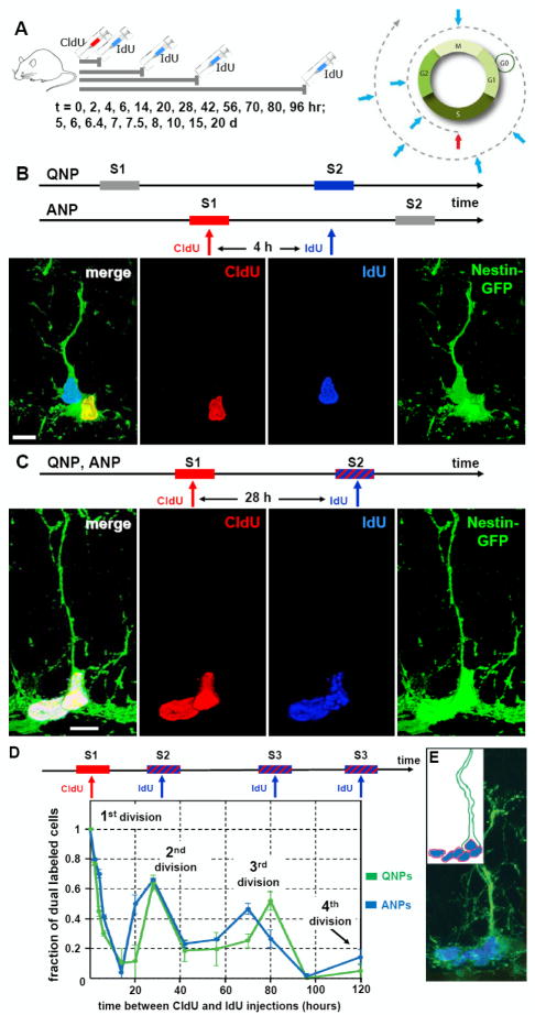
Fig. 6. Dynamics of QNP and ANP division in the DG.
(A) General scheme of the double S-phase labeling protocol. Nestin-GFP mice (2 months old, n=3 per time point) received a single injection of CldU followed, at different time interval (t), by a single injection of IdU. Scheme on the right illustrates that a cell marked by CldU injection during the S phase is able to incorporate the second label (IdU) if it is injected during the same S phase but not later, during the G2, M, or G1 phases. However, the cell can become double-labeled if IdU is injected when the CldU-labeled cell again enters the S phase during the second, third, and subsequent rounds of division.
(B) An example of double S-phase labeling with the CldU and IdU injections separated by 4 hr. In this example the interval between label injections is not commensurate with the length of the cell cycle of the ANPs or QNPs, and CldU labels the S-phase of the ANP, but not of QNP, whereas IdU labels the S-phase of the QNP, but not of ANP. On the micrograph the nucleus of the ANP cells is labeled with CldU (red) and the nucleus of the QNP cells is labeled with IdU (blue); co-localization of CldU and GFP is yellow.
(C) An example of double S-phase labeling with the CdU and IdU injections separated by 28 hr. CldU labels the S-phase of the first division cycle and IdU labels the S-phase of the second cycle (stripped bar). The length of the cell cycle is commensurate with the interval between label injections and some cells have incorporated both labels. On the micrograph the nuclei of a QNP and an ANP cells (distinguished by the presence of a GFP-positive radial process) are labeled with CldU (red) and IdU (blue); triple co-localization is white. Scale bar is 10μm in B, C.
(D) Distinct cycles of division can be detected by determining the fraction of CldU/IdU double-labeled QNP and ANP cells at given time intervals. The length of the cell cycles can be determined from the position of the peaks and the length of the S phase can be determined from the slope of the decrease during the first S phase. Both QNPs (green) and ANPs (blue) divide several times in a close succession as suggested by the presence of several peaks in dual labeling (the first peak corresponding to the 0 hr time point); note that the first division also reflects the asymmetric division of the QNP that gives rise to an ANP.
(E) A cluster of dividing cells in the SGZ. Several BrdU-labeled ANPs are located next to a BrdU-labeled QNP after 2 hr labeling; such clusters should be observed if QNP, as well as ANPs, reenters the cell cycle but not if it becomes quiescent after giving birth to an ANP (note that this experiment was performed with 6 month old mice, in which progenitor cells undergo a larger number of divisions than in 2 months old animals and in which there are fewer dividing cells such that the clusters of dividing cells are well separated). Inset: schematic representation of the cells and their BrdU-labeled nuclei.

