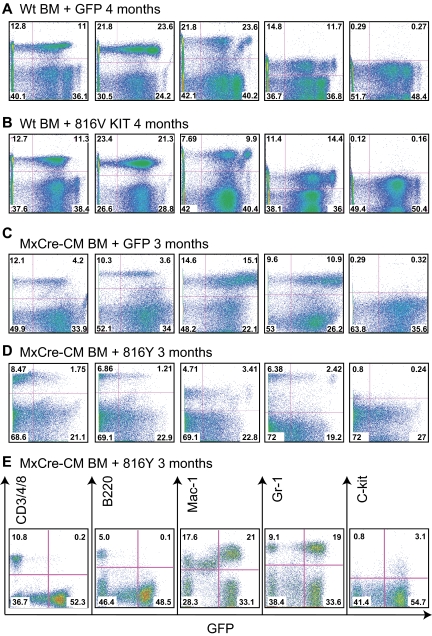
Figure 2.
Lineage contribution of KIT-transduced bone marrow cells in transplanted mice. PB cells were collected at the indicated times after transplantation from the recipient mice, stained for the indicated markers, and analyzed by FACS. (A) WT BM cells transduced with GFP. (B) WT BM cells transduced with KITD816. (C) Cbfb+/56m; Tg(Mx1-Cre) BM cells transduced with GFP. (D) Cbfb+/56m; Tg(Mx1-Cre) BM cells transduced with KITD816Y. (E) PB from a mouse that was starting to develop leukemia that was transplanted with Cbfb+/56m; Tg(Mx1-Cre) BM cells expressing KITD816Y. CD3/4/8 indicates combination staining of CD3, CD4, and CD8.

