Figure 4.
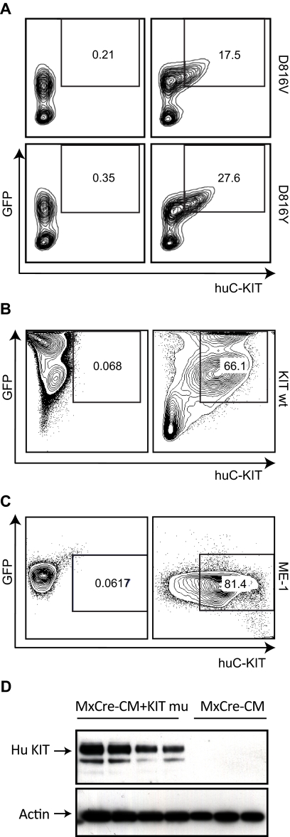
Expression of human KIT protein in the transplanted mice. (A) Leukemic cells from spleens of mice transplanted with Cbfb+/56m; Tg(Mx1-Cre)/KITD816V or Cbfb+/56m; Tg(Mx1-Cre)/KITD816Y BM cells. (B) Splenocytes from a mouse (nonleukemic) transplanted with Cbfb+/56m; Tg(Mx1-Cre)/KIT wt BM cells. (C) Leukemia cell line ME-1. The cells in panels A-C were stained with an anti–human KIT antibody and analyzed by FACS. In panels A through C, the cells in the left-hand panels were unstained and those in the right-hand panels were stained with the anti–human KIT. Cells in the boxes are GFP+ and human KIT+. (D) Western blot of leukemic spleen cells from Cbfb+/56m; Tg(Mx1-Cre)/KITD816Y (MxCre-CM + KIT mu) and Cbfb+/56m; Tg(Mx1-Cre) (MxCre-CM) mice using the anti–human KIT antibody. Actin was probed as the loading control.

