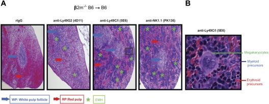
Figure 3.
Histologic evidence of BMC engraftment after Ly49G2 or Ly49C/I NK subset depletion of B6 recipients before β2m−/− BMC infusion. B6 recipient mice received mAbs for NK cell or NK-cell subset depletion using anti-NK1.1 (PK136), anti-Ly49C/I (5E6), anti-Ly49G2 (4D11) mAbs or rIgG control 2 and 1 day before lethal irradiation. A total of 15 million β2m−/− BMCs were transferred intravenously into B6 recipient mice. (A) H&E staining of formalin-fixed spleens (4×) for the different treatments after 7 days after transplantation is shown. (B) Enlarged image (40×) of a region that showed engraftment with marked EMH after anti–Ly49C/I (5E6) treatment. Blue and red arrows indicate the location of the white pulp follicle (WP) and red pulp (RP), respectively. Green asterisks indicate the EMH region. Images are representative of 2 independent experiments.

