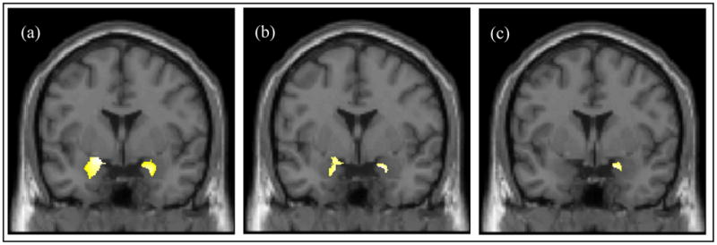
Figure 2.

(a) Significant reductions in gray matter concentration in BPD compared to HC; insets depict bilateral reductions in the medial temporal lobe. (Cluster peaks: Left Amygdala, t59 =5.06, x= −23, y= −2, z= −19; Right Amygdala, t59 =4.79, x= 23, y= −1, z= −22). (b) Significant reductions in gray matter concentration in female BPD compared to female HC. (Cluster peaks: Left Amygdala, t37 =3.94, x= −17, y= −0, z= −21; Right Amygdala, t37 =4.31, x= 20, y= 5, z= −19). (c) Significant reductions in gray matter concentration in sexually abused compared to non-abused female BPD subjects in Rt. medial temporal lobe. (Cluster peak: Right Amygdala, t18 =4.94, x= 18 y= 4, z= −19).
