Fig. 5.
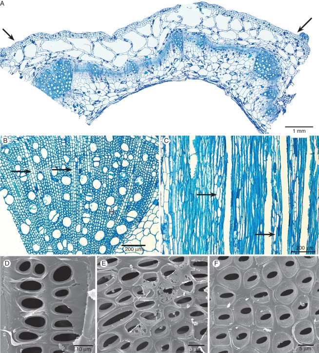
Stem anatomical pictures of Hydrocera triflora using light microscopy (A–C) and scanning electron microscopy (D–F). (A) Transverse section showing two stem ribs (arrows) and an aerenchyma region in the outer layer with large intercellular spaces. (B) Detail of transverse section showing wood formation at the rib region, vessels solitary or in small radial multiples, rays present (arrows). (C) Tangential section showing true rays with exclusively upright cells (arrows). (D) Tangential section near the pith showing enlarged intervessel pits with reduced pit borders. (E) Tangential section closer to the cambium; smaller intervessel pits with more pronounced pit borders. (F) Tangential section near the cambium; distinctly bordered intervessel pits in an opposite to alternate arrangement.

