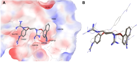
Fig. 10.
Binding poses of inhibitors modeled into the PC1/3 active site. A, molecule 166811 is shown in licorice; only polar hydrogen bonds are displayed. The molecular surface of PC1/3 binding site is colored by electrostatic potential. B, overlay of docking poses obtained for four PC1/3 inhibitors.

