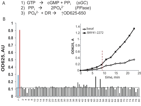
Fig. 1.
Screening for sGC activators. A, the principle of the coupled sGC/pyrophosphatase assay: PO42− ion produced after the conversion of GTP into cGMP and phosphate (reactions 1 and 2) is quantified by the reaction with a malachite green/molybdate mixture (DR) and detected by absorbance at 630 nm (reaction 3). B, colorimetric detection of PO42− produced in the assay after incubation with different library pharmacophores. The reaction mixture was pretreated with 100 μM pharmacophores from the ChemBridge library (■) before addition of 200 μM GTP. Representative data from one screening plate with 80 pharmacophores are shown. The values were compared with control samples (C) in which sGC was treated with the vehicle (□), 5 μM BAY41-2272 (blue bar), or a mixture of 10 μM ODQ and 100 nM BAY58-2667 (red bar). Inset, the assay is linear over a broad time range, and the difference between basal and BAY41-2272-stimulated sGC activity is significant after 9 min (vertical line). Data are means ± S.D. of two independent measurements performed in duplicate. PPi, inorganic pyrophosphatase; AU, absorbance unit.

