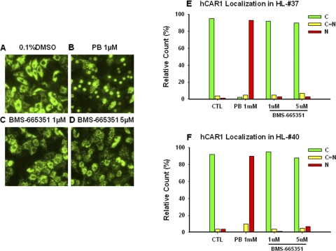
Fig. 4.
Nuclear translocation of hCAR in human primary hepatocytes. Human primary hepatocytes from donors HL-37 and HL-40 were infected with Ad/EYFP-hCAR as outlined under Materials and Methods. Twelve hours after infection, HPHs were treated with vehicle control (0.1% DMSO), PB (1 mM), or BMS-665351 (1 and 5 μM) for 8 h. Subsequently, cellular localizations of infected hCAR were visualized under a confocal microscope. A to D, representative Ad-EYFP-hCAR localizations from vehicle control, PB-treated, and BMS-665351-treated hepatocytes (donor HL-37). E and F, 100 Ad/EYFP-hCAR expressing HPHs from each treatment group were calculated with a percentage of cytosol, nucleus, and mixed (cytosol + nucleus) localizations for donors HL-37 (E) and HL-40 (F).

