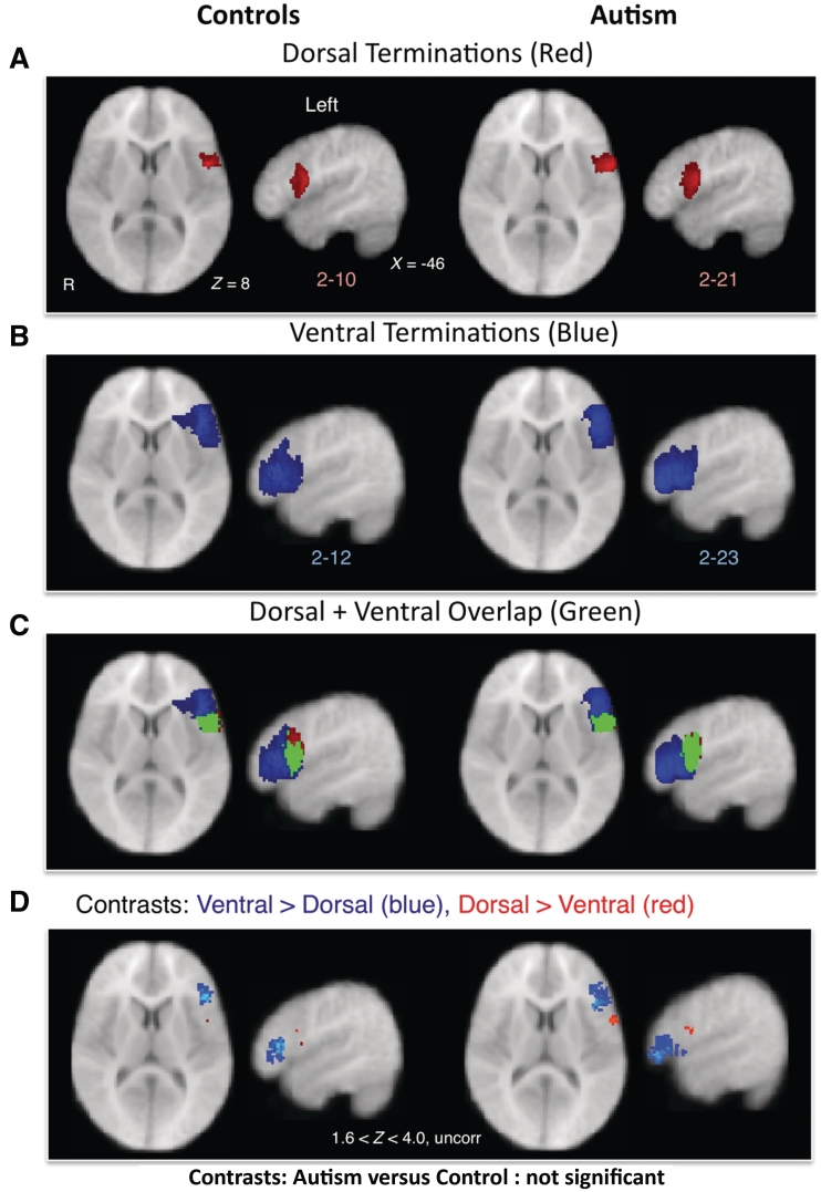
Figure 5.
DTI tractography of dorsal and ventral language pathways. (A–C) Termination points (with a minimum overlap of two subjects for visualization purposes) of left dorsal (A, red) and ventral (B, blue) pathways in control (left) and autistic (right) subjects as determined by DTI tractography from A1 to inferior frontal gyrus. (C) Green represents areas that overlapped for both dorsal and ventral terminations. (D) Within-group comparisons between proportions of subjects with dorsal and ventral terminations in inferior frontal gyrus. In both control and autistic subjects, ventral > dorsal terminations (blue) were more anterior and dorsal > ventral terminations (red) were more posterior (Z > 1.6, P < 0.05, uncorrected). Analysis failed to confirm differences in terminations of either tract for control versus autistic comparisons. R = right.

