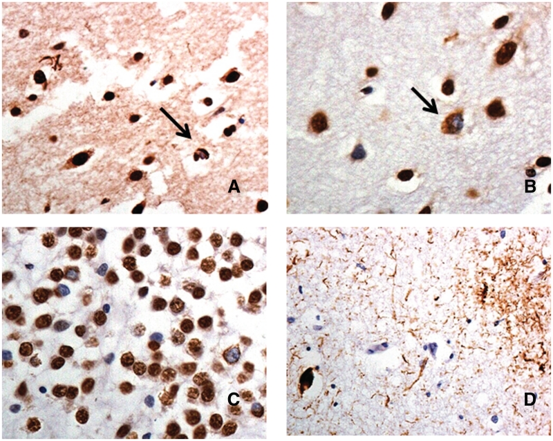
Figure 2.
Immunohistochemical staining for TDP-43 and tau proteins in patients bearing the hexanucleotide repeat expansion in C9ORF72. Patient 10 shows occasional TDP-43 immunoreactive neuronal cytoplasmic inclusions (arrow) and dystrophic neurites in the outer layers of the temporal cortex consistent with FTLD-TDP type A histology (A). Patient 6 shows occasional, often granular, TDP-43 immunoreactive neuronal cytoplasmic inclusions (arrow) in the outer layers of the temporal cortex (B), and many granular inclusions in dentate gyrus granule cells (C), consistent with FTLD-TDP type B histology. Patient 24 shows a tauopathy with astrocytic plaque and neurofibrillary tangle-like structures, consistent with corticobasal degeneration (D). Immunoperoxidase—haematoxylin, magnification ×40.

