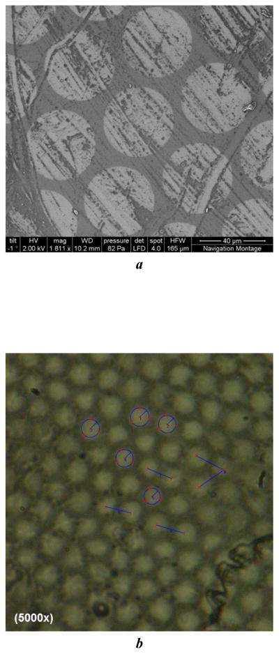
Fig. 1.

Sample fiber bundle images: (a) Scanning Electron Microscope (SEM) images for type I fiber bundle (core spacing: 50μm); (b) Digital microscope image for type III fiber bundle (core spacing: 4.5μm).

Sample fiber bundle images: (a) Scanning Electron Microscope (SEM) images for type I fiber bundle (core spacing: 50μm); (b) Digital microscope image for type III fiber bundle (core spacing: 4.5μm).