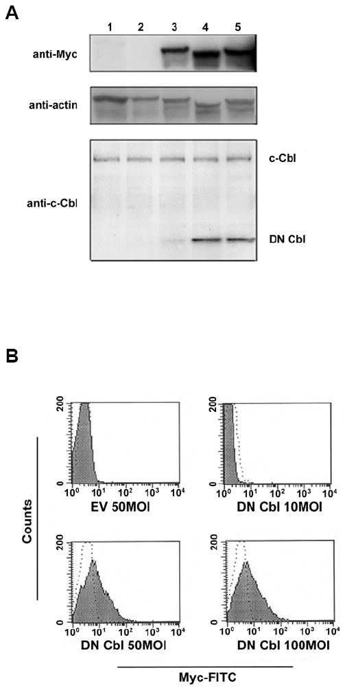
Figure 1. DN Cbl protein is expressed in CD4+ T cells after Ad transduction.

CAR Tg Th1 T cells were transduced with EV or Ad-DN Cbl and cultured overnight. On the next day, cells were analyzed for DN Cbl expression. (A) Representative image of a Western blot assay. Lane 1: Non-transduced cells. Lane 2: EV-transduced CAR Tg Th1 T cells. Lanes 3-5: Ad-DN Cbl-transduced CAR Tg Th1 T cells at an MOI of 10, 50, or 100. The upper panel was blotted with anti-myc tag antibody, the medium panel with anti-β-actin antibody as a loading control, and the lower panel with anti-c-Cbl N-terminus antibody. (B) Intracellular staining assay. Transduced CAR Tg Th1 T cells were fixed, permeabilized, and stained with a FITC-conjugated anti-myc tag antibody as shown in the solid area or isotype control antibody as shown in dotted line.
