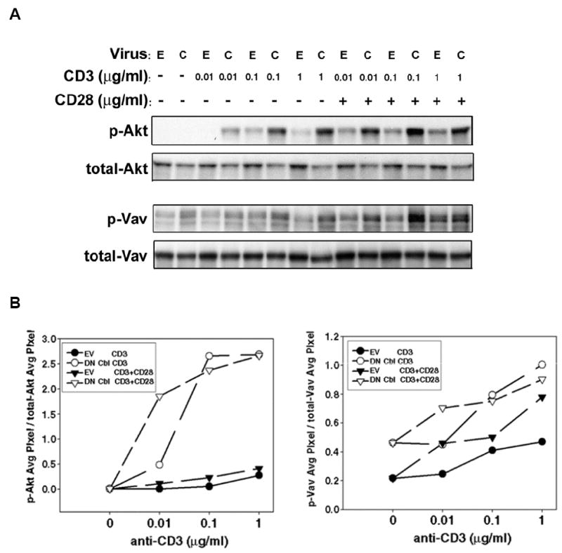
Figure 3. DN Cbl predominately augments a TCR signal.

(A) CAR Tg Th1 T cells were transduced with EV or Ad-DN Cbl. On the next day, transduced cells were stimulated with serially titrated anti-CD3 antibody-coated beads with or without the presence of 1μg/ml anti-CD28 antibody on the beads, for 15 min. Cells were then lysed for Western blotting for phospho- and total-AKT (upper panels) and phosho- and total-Vav (lower panels). (B) The blots were analyzed using UN-SCAN-IT software for quantitation of the phosphorylated band intensities relative to the total amount of the protein density detected.
