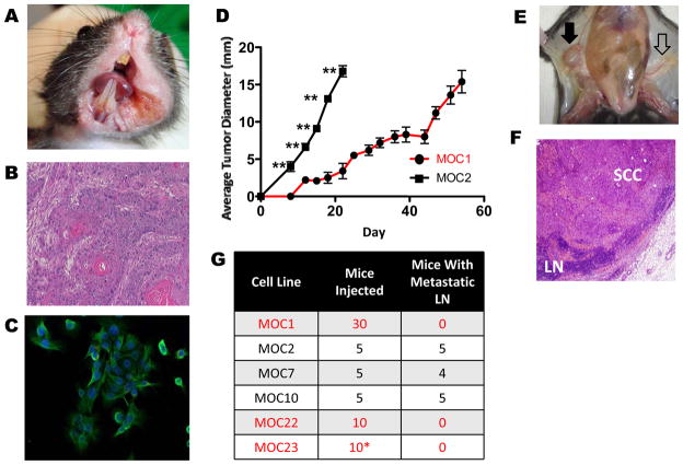
Figure 1. Variant growth patterns and lymphatic metastasis in a C57BL/6 syngeneic model of OSCC.
A. Representative primary OSCC in left floor of mouth/buccal region after 25 weeks of biweekly DMBA treatment (MOC10 parent tumor). B. Representative H&E stained section of primary tumor (MOC10 parent tumor) showing moderately differentiated squamous cell carcinoma (40X magnification). C. Immunofluorescence of MOC10 cell line showing epithelial phenotype with positive cytokeratin staining (green) and DAPI for nuclear staining (40X magnification). D. Representative in vivo growth curves comparing indolent MOC1 and aggressive MOC2 after 1×10⁶ cells were injected into the right flank of C57BL/6 WT mice (**=p<0.01 in all days post day 0). E. Metastatic inguinal draining lymph node (black filled arrow) that is enlarged and discolored compared to contralateral normal appearing lymph node (black open arrow). F. H&E stained metastatic lymph node shows effacement of normal architecture by SCC (LN= lymph node, 20X magnification). G. Summary of LN metastatic capacity of each cell line. (*MOC23 data is shown for RAG2−/− mice, as this line does not form progressive tumors in WT mice)

