
Figure 2. Expression of heparanase in non-TUNEL-positive cells.
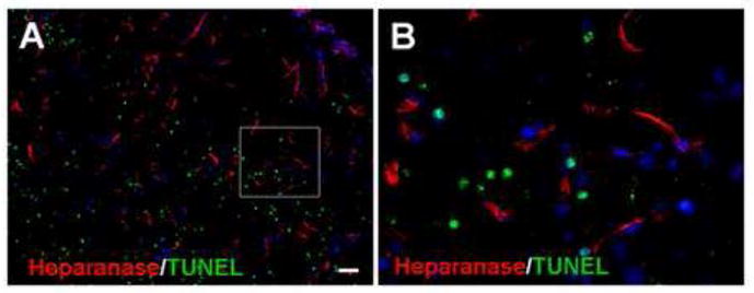
Immunostaining revealed heparanase expression (red) in the peri-infarct region 7 days after ischemia. TUNEL staining (green) was applied to detect injured and dead cells. The double staining images in A and B show that there was little overlap between heparanase and TUNEL staining, suggesting that heparanase-positive cells were not injured or dead cells. Blue is nuclei staining with Hoechst 33258. Bar: A=50μm, B=10μm.