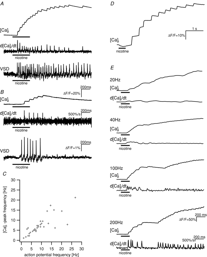
Figure 1. Patterns of [Ca2+]i peaks and action potential discharge showed a high degree of similarity in individual neurones.

A and B show the response patterns to the application of nicotine (100 μm, horizontal bars) for the [Ca2+]i signal (top traces, scale bar in B), the first derivative of the calcium signals (middle traces, scale bar in B) and the change in membrane potential (bottom traces, scale bar in B) for two neurones. [Ca2+]i signals and changes in membrane potential were measured consecutively. Similarities between [Ca2+]i peaks and action potential discharge can be seen for long- (A) and short- (B) lasting spike discharge. C, frequency of action potentials and [Ca2+]i peaks is highly correlated in cultured myenteric neurones (Spearman's rank order correlation, P < 0.001). The frequencies were determined in separate, consecutive measurements of changes of membrane potential and [Ca2+]i in response to the application of nicotine in 34 neurones. D, change of [Ca2+]i in response to the application of nicotine in a neurone of a whole-mount preparation from the human submucous plexus. The signal shows the same characteristics as the signals from cultured guinea-pig myenteric neurones. E, nicotine evoked [Ca2+]i signals in a single neurone of guinea-pig myenteric plexus. While frame rates of 200 Hz allowed the detection of fast [Ca2+]i peaks, the lower frame rates of 20, 40 or 100 Hz only revealed slow [Ca2+]i transient or did not fully resolve the [Ca2+]i peaks, respectively. The first derivative of the calcium signals are shown below the [Ca2+]i traces.
