
Figure 4. Stimulation of murine corticotrophs by CRH/AVP.
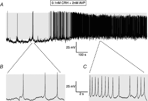
A, representative current clamp recording of a metabolically intact corticotroph before and following exposure to a 3 min pulse of 0.1 nm CRH and 2 nm AVP. B and C, expanded traces before application of CRH/AVP (B) and immediately following washout of secretagogue (C). Grey shading indicates membrane potential between −50 and +10 mV.