
Figure 1. mCCDC11 cells release exosomes.
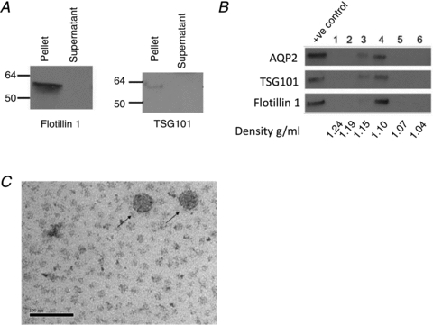
A, in the mCCDC11 cell culture medium, the exosomal marker proteins flotillin-1 and TSG101 localise to the ultracentrifugation pellet (pellet) rather than the supernatant. B, Western blot for aquaporin 2 (AQP2), flotillin-1 and TSG101 on fractions obtained following isopycnic centrifugation. The exosomal markers are present in fractions corresponding to a density of 1.10–1.15 g ml-1. The positive control was unfractioned exosomes. C, structures of exosome size and shape are visible in the cell culture medium using transmission electron microscopy. Arrows indicate 2 exosomes. Bar, 100 nm.