
Figure 3. mCCDC11 cells exposed to AQP2-containing exosomes express functional AQP2.
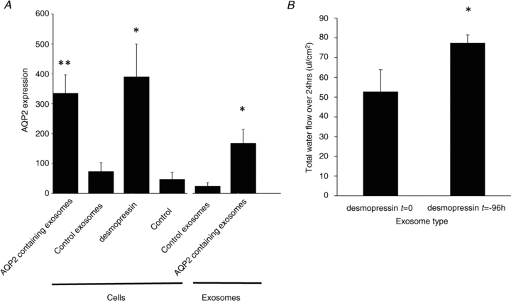
A, AQP2 protein expression was measured by Western blot and band densitometry. Cellular AQP2 expression (‘cells’) was measured after 48 h incubation with AQP2-containing exosomes (derived from mCCDC11cells treated with desmopressin (3.16 ng ml-1) for 96 h); control exosomes derived from unstimulated cells or direct treatment with desmopressin (3.16 ng ml-1). The concentration of AQP2 in the exosomes is also presented (‘exosomes’). **p < 0.02 vs. control. *p < 0.05 vs. control. n = 4. B, the water flow across mCCDC11monolayers after 48 h co-culture with exosomes from mCCDC11cells exposed to desmopressin (3.16 ng ml-1) immediately before exosome isolation (desmopressin t = 0) or 96 h before exosome isolation (desmopressin t = –96 h). Water flow was determined by weighing the medium in the apical and basolateral compartments. Water flow is expressed as μl per area of monolayer. *P = 0.05 by paired analysis. n = 4.