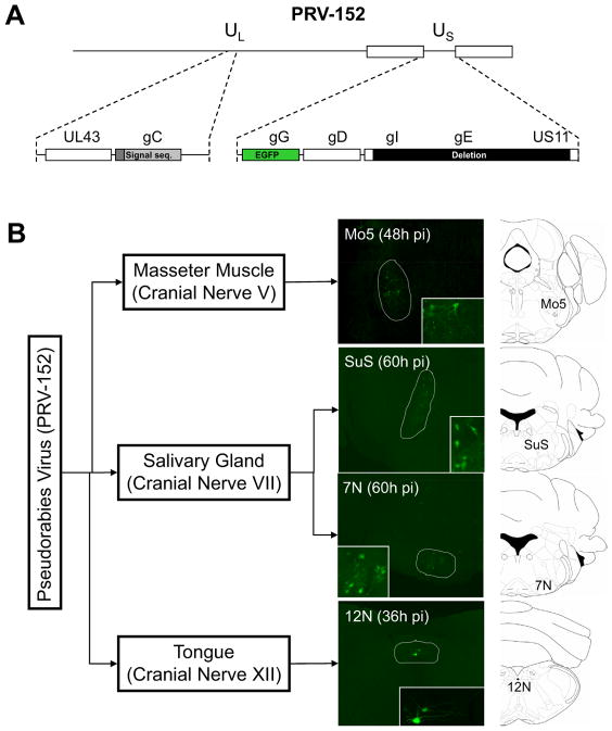
Figure 1. Brainstem Cranial Nerve Motor Nuclei are the First CNS Infected Targets Following PRV Injection Into Peripheral Tissues.
A) PRV-152, a Bartha PRV strain genetically modified to express EGFP constitutively (Smith et al., 2000) was used in the present studies. B) PRV-152 was injected bilaterally into the submandibular salivary gland (SAL), the masseter muscle (MAS), or unilaterally into the posterior part of the tongue (TP). 36, 48 or 60 hrs after the injections, EGFP immunofluorescence was analyzed in the brains of infected animals. The motor nucleus for the indicated cranial nerves was the first region where PRV infection was detected, following PRV injection into the indicated tissues. The motor nucleus of the cranial nerve V (Mo5) was the first brainstem area infected after PRV-152 injection into the masseter muscle (MAS), the motor nucleus of the cranial nerve VII (7N) and the superior salivatory nucleus (SuS) were identified after PRV injection into the salivary gland (SAL). Finally, the motor nucleus of the cranial nerve XII (12N) was the only place where PRV was readily observed after PRV injection into the posterior region of the tongue (TP).

