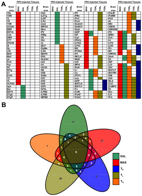
Figure 2. Brain Areas Infected by PRV After Injection Into Multiple Peripheral Tissues.
A) Distribution of forebrain areas infected by PRV-152 96hr after five distinct PRV injections (SAL, MAS, TP, TL, TA) were made into peripheral tissues. This analysis revealed that several brain areas were infected by PRV in each circuit studied (color- coded columns). Very few brain regions were common to the descending neural circuits projecting to all peripheral tissues injected. These regions included the lateral hypothalamus, basolateral and central amygdala and insular, ectorhinal and perirhinal cortices (see multiple color-coded rows in the lower-right corner). B) Symmetrical, non-proportional, 5-way Venn diagram illustrating the number of PRV-infected brain regions after injection in 1 to 5 peripheral sites. It can be appreciated the vast number of subsets (32) to be considered when analyzing multiple circuits and how our proposed approach can identify brain regions belonging to only one such subset. n=3 per injection site.

