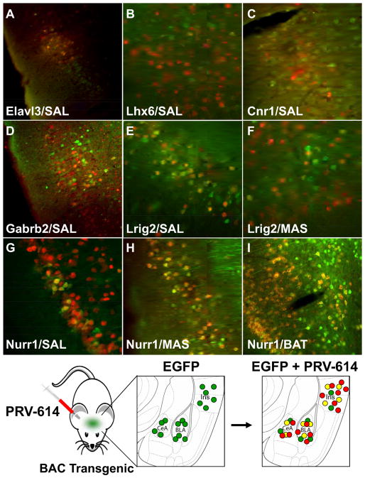
Figure 4. Colocalization of PRV-Encoded mRFP and Transgenic EGFP Reveals Markers for Neurons in Feeding Neuronal Circuits.
Elavl3+/PRV+ neurons (A) were detected in the rhinal cortex following a SAL PRV injection. Lhx6+/PRV+ neurons (B) were detected in the insular cortex after a MAS PRV injection. Cnr1+/PRV+ neurons (C) were detected in the insular cortex following a PRV injection into SAL. ; Gabrb2+/PRV+ neurons (D) were detected in the insular cortex following a PRV injection into SAL. Lrig2+/PRV+ neurons were detected in the insular cortex following PRV injections into SAL (E) and MAS (F). Nurr1+/PRV+ neurons were detected in the insular cortex following SAL (G), MAS (H) and Brown Adipose Tissue (I) PRV injections. (J) Diagram showing our approach to identify markers for neuronal populations from a specific neural circuit (as indicated by their susceptibility to PRV infection). Mice in which EGFP is known to be expressed in a specific brain region are infected in the periphery with PRV-614 (encodes mRFP) and in those cases where both mRFP (from PRV) and EGFP (from the transgenic mouse) are detected in the same neuron, it can be asserted that the gene whose promoter directs EGFP expression is a marker for neurons integrated into the circuit projecting to the tissue infected by PRV-614.

