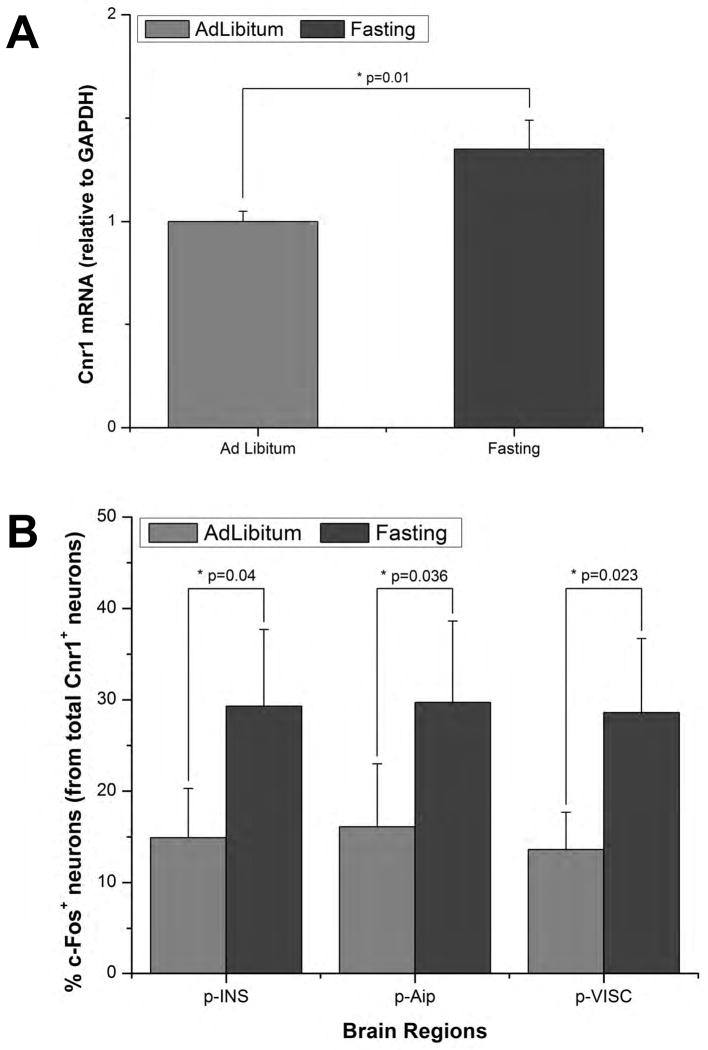
Figure 7. Insular Cnr1+ Neurons are Sensitive to Fasting.
(A) Insular cortex Cnr1 mRNA levels were measured in wild type mice fed ad libitum vs. mice that had fasted for 36 hrs. Using a Taqman assay it was determined that there was a significant increase in Cnr1 mRNA following a 36h fasting period (p=0.01, n=5). (B) We also quantified the number of c-Fos+/Cnr1+ neurons in the insular cortex of Cnr1::EGFP mice in mice with unrestricted access to food vs. mice that had fasted for 36hrs (n=3 per group). It was observed that the number of c-Fos+/Cnr1+ neurons in the posterior insular cortex (p-INS) doubled following fasting (p=0.040); this was also valid for its agranular (p-Aip; p=0.036) and visceral (p-VISC; p=0.023) subdivisions. All data are represented as mean +/- SEM; Student's t test was used for statistical analysis. Significance was assumed for p values < 0.05.

