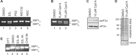
Figure 2. Inhibition of translocation promotes ER stress.
(A) HeLa cells were incubated with DMSO, 8 μM ESI, 10 μM PS2, 10 μM MG132 or 0.4 μM of the deubiquitinase inhibitor NSC687852 (NSC) for 8 h. Xbp1 mRNA splicing was determined by RT–PCR. Products corresponding to unspliced (u) and spliced (s) Xbp1 mRNA are indicated. (B) HeLa cells were incubated with DMSO or the indicated concentration of the translocation inhibitor cpd A for 8 h, and Xbp1 splicing was determined as above. (C) HeLa cells were incubated with DMSO or 10 μM cpd A for 8 h, and lysates were analysed by blotting with anti-peIF2α (phospho-eIF2α) or anti-eIF2α antibodies. (D) HeLa cells were incubated with DMSO or 10 μM cpd A for 8 h, pulse-labelled with [35S]Met/Cys for 40 min, and lysates were analysed by phosphorimaging (top panel) or blotting with an anti-tubulin antibody (bottom panel). Molecular mass in kDa is indicated on the left-hand side. (E) HeLa cells were incubated with 2 mM DTT for 2 h, DMSO for 8 h, 8 μM ESI for 4 or 8 h, or 100 μg/ml CHX for 8 h. Xbp1 splicing was determined as above.

