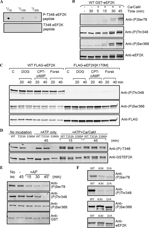
Figure 5. Use of phosphospecific antibodies to study the autophosphorylation of eEF2K in vitro and in intact cells.
(A) Dot blot to establish whether the anti-phospho-Thr348 antibody is truly phosphospecific. A total of 1 μl of the indicated dilutions of the phosphorylated and non-phosphorylated versions of the corresponding peptide (10 mg/ml) were applied to the membrane, which was developed as for Western blot analysis. (B) Recombinant GST–eEF2K expressed in E. coli was incubated without or with Ca2+/CaM, as indicated, and non-radioactive ATP for the times shown. Samples were then analysed by SDS/PAGE followed by Western blot analysis with the indicated antibodies specific for phospho (P)-Ser78, phospho-Ser366 or phospho-Thr348, and anti-eEF2K antibody as a loading control. (C) Results for the phosphorylation of Thr348 and Ser366 in vivo. HEK-293 cells were transfected with a vector encoding either wild-type (WT) eEF2K or the kinase-defective eEF2K[K170M] mutant. Cells were treated for the indicated times with 2-deoxyglucose (DOG; 10 mM; after transfer to medium containing 5 mM glucose), chlorophenylthio-cAMP (CPT-cAMP; 0.5 mM) or forskolin (Forsk; 10 μM); the latter two were used in the presence of isobutylmethylxanthine (1 mM). Cell lysates were analysed by SDS/PAGE and immunoblotting using the indicated antibodies. C, control (untreated) cells. (D) Recombinant GST–eEF2K was incubated, where noted, with non-radioactive MgATP and, in some cases, Ca2+/CaM for the indicated times. Samples were then denatured and analysed by SDS/PAGE and Western blotting using the indicated antibodies. (E) Recombinant GST–eEF2K was either analysed directly (‘No inc’) or incubated with alkaline phosphatase (+AP) for the times shown. Samples were analysed by SDS/PAGE and Western blotting using the indicated antibodies. (F) Wild-type eEF2K or two kinase-deficient mutants (K/M, eEF2K[K170M]; D/A, eEF2K[D274A]) were analysed by SDS/PAGE and Western blotting using the indicated antibodies.

