Fig. 1.
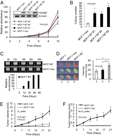
Effect of HOXB7 expression in breast cancer cells. (A and B) (A) Immunoblot analysis of HOXB7 expression in MCF-7-Vec and MCF-7-B7 cells (three clones: MCF-7-B7 1, 2, and 3) and growth curve of MCF-7-Vec and MCF-7-B7 cells grown in monolayer culture and (B) soft agar colony formation by MCF-7-Vec and MCF-7-B7 cells. (C) T1-weighted 1H MR imaging of MCF-7-B7 cells, and MCF-7-Vec cells visualizing the degradation of ECM over a period as indicated. (D) Representative 3D reconstructed images of vascular volume maps (row 1), permeability-surface area product (row 2), and combined vascular volume and permeability-surface area product (row 3) for MCF-7 parental (column 1), MCF-7-Vec (column 2), and MCF-7-B7 (column 3) tumors in mice. (E) Tumor growth curves of MCF-7-Vec and MCF-7-B7 cells implanted s.c. in athymic mice in the presence of an exogenous slow release, estrogen implant and (F) in the absence of exogenous estrogen supplementation.

