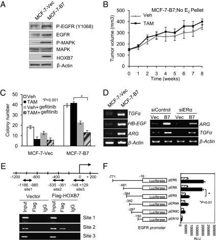
Fig. 3.
HOXB7 promotes TAM resistance. (A) Immunoblot analysis of Phospho-EGFR, EGFR, P-MAPK, MAPK, and HOXB7 expression levels in MCF-7-Vec and MCF-7-HOXB7 cells. (B) Tumor growth curve of MCF-7-HOXB7 cells implanted s.c. in athymic Swiss female mice and treated with either vehicle or TAM (83.3 μg/d), in the absence of an exogenous estrogen supplement. (C) Soft agar colony formation by MCF-7-Vec and MCF-7-B7 cells treated with vehicle, estrogen (10 nM), or TAM (1 μM) and combination with 1 μM gefitinib as indicated. (D) Semiquantitative RT-PCR analysis of mRNA expression levels of TGF-α, HB-EGF, and Amphiregulin in HOXB7-expressing MCF-7 cells and their vector controls (Left) and Amphiregulin or TGF-α mRNA expression in MCF-7-Vec and MCF-7-B7 cells treated with either scrambled sequence siRNA or ERα-specific siRNA (Right). (E) Diagram representing the HOXB7 binding sites in the EGFR promoter region. MCF-7 cells were transfected with pcDNA3-Flag-HOXB7 and vector plasmid, and ChIP was performed by IP with either anti-Flag M2 antibody or control IgG. (F) Luciferase activity of deletion/truncation constructs of the EGFR promoter, with (solid bars) and without (hatched bars) transfected HOXB7 plasmid, to map the minimal region necessary for activation by HOXB7.

