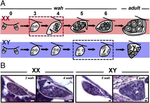
Fig. 2.
Time course of gonadal sex differentiation in XX and XY fish. (A) Schematic representation of the morphological changes in female and male gonads during sex differentiation (wah: weeks after hatching). (B) Light histology of the critical developmental stages indicated by dotted boxes in A. Black (XX; 4 wah) and white (XY; 6 wah) arrowheads indicate the appearance of somatic cell outgrowths (ovarian differentiation) and rudiments of the main sperm duct (testicular differentiation), respectively.

