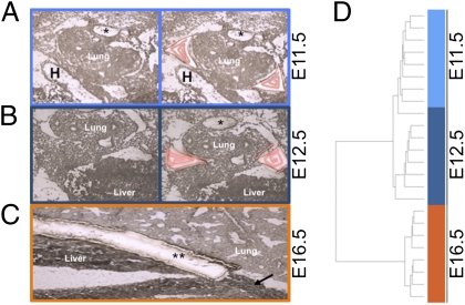
Fig. 1.
Expression study of microdissected tissue. (A and B) Unstained and unfixed transverse cryosections of E11.5 (A) and E12.5 (B) mouse embryo before (Left) and after (Right) laser capture microdissection (LCM) (H, heart; *, fused dorsal aorta). Captured tissue is outlined in red. (C) Sagittal cryosection of an E16.5 mouse embryo showing an anatomically mature diaphragm (arrow) (**, microdissected diaphragm). (D) Unsupervised hierarchical clustering of 871 differentially expressed probes (Welch modified t test) identifies two distinct clusters [the early (E11.5, E12.5) and late (E16.5)] Additionally, E11.5 and E12.5 separate in two subclusters, except for one outlier.

