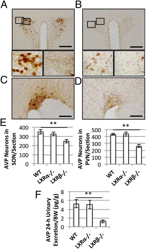
Fig. 4.
Immunohistochemical detection of hypothalamic vasopressin-positive neurons. (A) Representative hypothalamic section of the PVN of a 12-mo-old WT mouse. (Scale bar: 100 μm.) (Bottom) Higher magnification of the selected areas showing AVP-positive cell bodies (Left) and neuronal projections (Right). (B) PVN of a 12-mo-old LXRβ−/− mouse. (Scale bar: 100 μm.) (Bottom) Higher magnification of selected areas. Reduced immunoreactivity for AVP was detected both in the magnocellular neurons (Left) and in their projections (Right). (C and D) Representative sections of SON in WT (C) and LXRβ−/− mice (D). (Scale bars: 50 μm.) (E) Numbers of AVP-positive neurons per section in SON (Left Inset) and PVN (Right Inset). Data are presented as mean ± SEM; **P < 0.001 versus WT. n = 6 in each group. (F) Urinary AVP excretion in 24 h. The urinary AVP content was reduced significantly in LXRβ−/− mice compared with WT animals. Data are presented as mean ± SEM; **P < 0.001 versus WT. n = 6 in each group.

