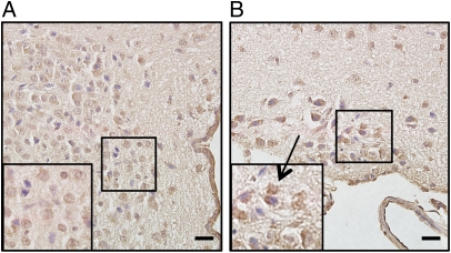
Fig. 5.
Immunohistochemical study of the expression of LXRβ in the hypothalamus of 8-mo-old WT female mice. (A) A representative section of the PVN. Positive immunoreactivity for LXRβ is detectable in the magnocellular neurons. (B) Positive staining for LXRβ is shown in the SON (arrow). Insets show higher magnification of the selected areas. (Scale bars: 20 μm.)

