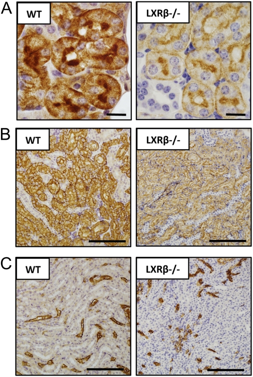
Fig. 6.
Immunohistochemical study of AQP-1 expression in the kidney of 12-mo-old WT and LXRβ−/− female mice. In WT mice AQP-1 immunoreactivity was detectable in the renal proximal tubule (A), in the descending thin limb of Henle (B), and in the vasa recta (C). In LXRβ−/− mice the positive staining was evident with a markedly lower reactivity in the proximal tubule (A) and in the descending thin limb of Henle (B) but not in the vasa recta (C). (Scale bars: 10 μm in A; 100 μm in B and C.)

