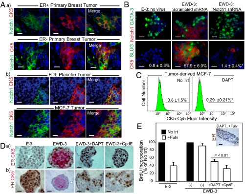
Fig. 4.
Expansion of luminobasal cells by EWD involves Notch signaling. (A) Paraffin sections of (a) primary breast cancers or (b) E-3 and MCF-7 xenografts were dual-stained for Notch1 (green) and CK5 (red). (Scale bars, 20 μm.) (B) EWD-3 cells were transduced with lentivirus encoding an shRNA targeting Notch1 or a scrambled control shRNA and propagated for >45 d under EWD conditions. Paraffin sections of 3D colonies were stained by dual IHC for CK5, Notch1, Slug, and GATA-3 for comparison with luminal E-3 colonies. Numbers are percentage luminobasal content. *P < 0.01. (C) Flow analysis of CK5 expression in tumor-isolated MCF-7 cells treated 14 d with 2 μM DAPT. *P < 0.01. (D) Line 3 was grown >45 d in E or EWD media, or EWD supplemented with the GSIs DAPT or CpdE. 3D colonies were stained by IHC for CK5 (red) and (a) ER or (b) PR (brown). (E) Colonies grown as in D were treated 7 d with 100 nM Fulv. Cell proliferation was assessed by IHC staining for BrdU-positive nuclei. Inset: IHC for ER. Receptors are degraded by Fulv.

