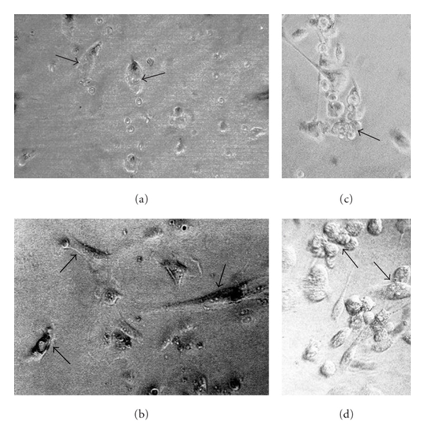
Figure 4.

Examples of in vitro culture of primary human cancer cells derived from surgical biopsies. (a) and (c) Primary ovary cystadenocarcinoma-derived cells. (b) and (d) Primary prostate adenocarcinoma-derived cells. Parental ovary cancer cells (a) and prostate cancer cells (b) are attached efficiently in the fourth day of culture (arrows). “Antisense/triple helix” anti-IGF-I ovary cancer cells (c) and prostate cancer cells (d), both twenty days after transfection, form the established lines characterized often by the clusters of round apoptotic cells, becoming progressively small (c, arrow; d, arrow up). They are accompanied by nonapoptotic and more voluminous cells (d, arrow down) presenting generally elongated shape (c and d). The transfected cells are always different from nontransfected parental cells (a, b), as it was demonstrated previously in cases of human glioma and hepatoma cell lines established from primary tumours of glioblastoma and hepatocarcinoma [30, 31] X400.
