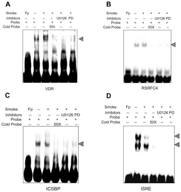
Fig. 4.
Effects of ERK 1/2 inhibitors on TS-regulated DNA binding activity in A549 cells. Cells were treated with and without TS and nuclear proteins were extracted and incubated with biotin-labeled probes corresponding to different TF binding sequences for EMSA as described in MATERIALS AND METHODS. A: VDR. B: RSRFC4. C: ISCSP. D: ISRE. Lane 1 of each panel indicates free probes. DNA binding activity is represented without (lane 2) and with (lane 3) TS exposure in each panel. Lane 4 of each panel represents binding activity after TS exposure but with 20× excess of cold probes added to the incubation. Lane 5 (U0126) and lane 6 (PD98059) are TS-exposed binding in the presence of ERK 1/2 inhibitors. These results are representative of 3 independent experiments. Fp, free probe.

