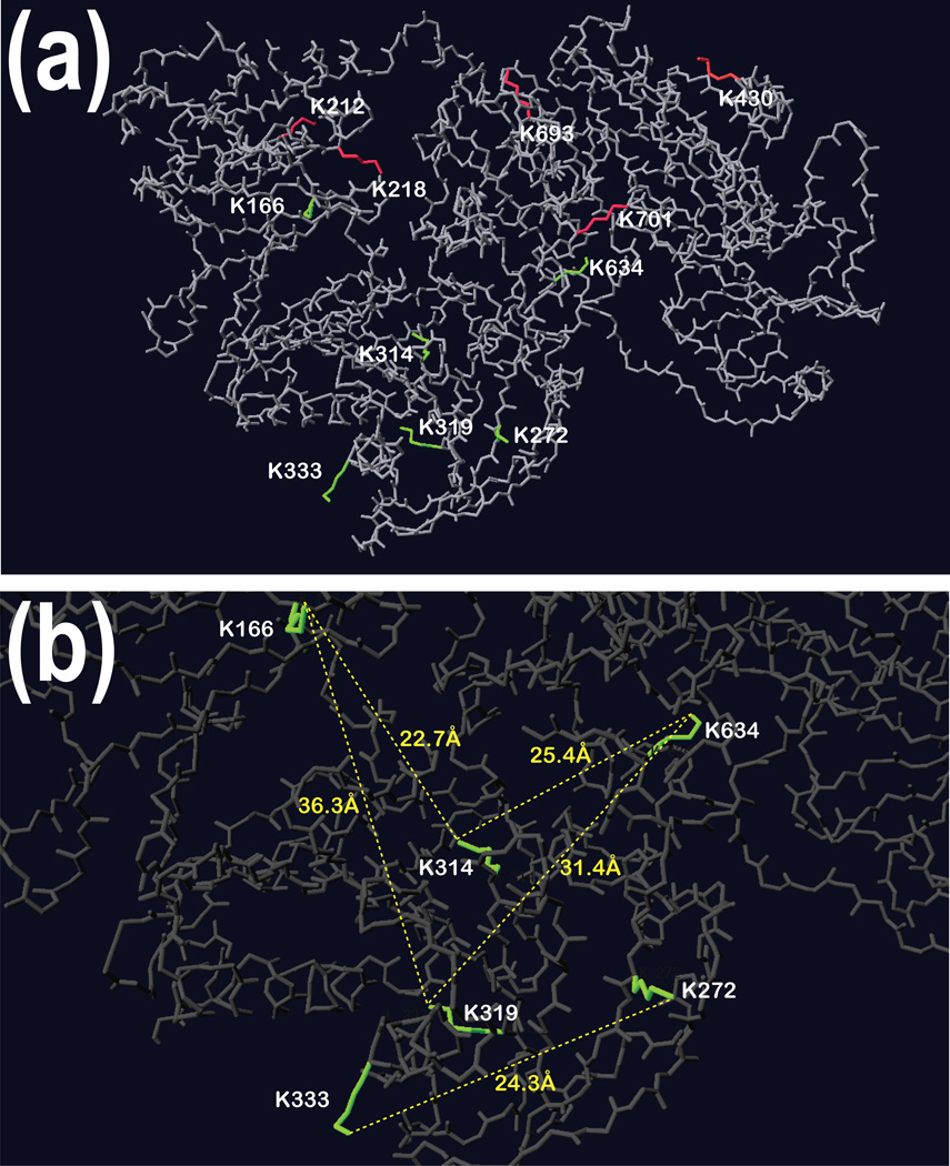
Figure 6. An overlay of identified modified lysine residues on 3-dimensional structure of GSN.
Panels show the analysis of the modified lysines on the 3D structure of gelsolin. Panel a: Red labeled Lys residues linked together within the same peptide, as well as the dead-end modifications (Lys 420) are located on the surface of gelsolin. Green labeled Lys residues involved in the cross-linking of the dimer of gelsolin are mostly found in cavities of the protein. Pane b: The distances between the ε amine of Lys residues involved in the dimerization reveal a distance that is longer for the cross-linker to reach both amines.

