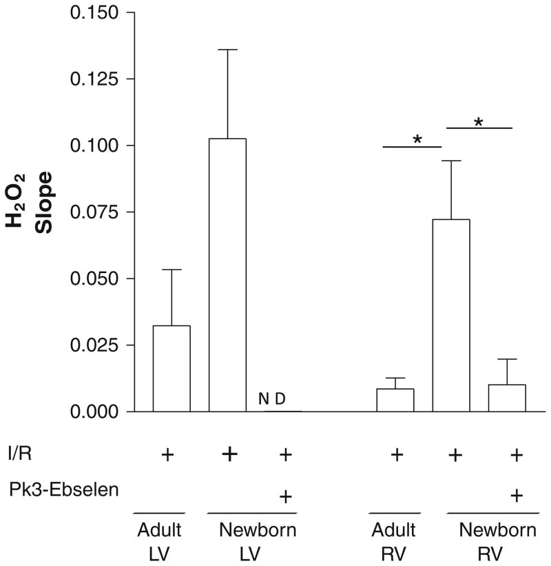
Fig. 6.
H2O2 is increased in the right ventricle of newborns compared with that of adults after hypoxia. H2O2 generation in newborn and adult LV and RV myocytes was measured using real-time confocal microscopy on isolated myocytes (after incubation with DCFDA) after 90 min of IR. A fivefold increase was observed in the newborn right ventricle compared with adult RV cells (*P < 0.05 [ANOVA followed by Bonferroni posttest]), whereas no significant change was found in the newborn left ventricle compared with that of the adult. A threefold decrease was observed in the newborn right ventricle, whereas no signal was detected in the newborn left ventricle after PK3-ebselen treatment. Values are mean ± SE; n = 6 to 8 cells per group. ND not detected

