
Figure 4.
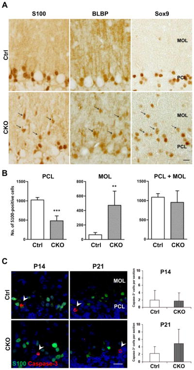
Translocation of Bergmann glial cell bodies into the MOL in APC-CKO mice. A. The cells positive for Bergmann glial markers S100, BLBP, and Sox9 are predominantly localized in the PCL and only a few are found in the MOL of control mice (Ctrl) at P21. In contrast, a number of Bergmann glial markers-positive cells are distributed within the MOL of CKO mice at the same age (arrow). B. Quantification of S100+ cells in the PCL and MOL. The number of S100+ cells is significantly lower in the PCL but significantly higher in the MOL of CKO mice compared to those of Ctrl, whereas the total number of S100+ cells in the MOL and PCL is not significantly different. n=5, **p<0.01, ***p<0.001 versus controls, t test. C. No increase in glial cell apoptosis in the cerebellar cortex of CKO mice. Representative images and quantification of caspase-3+ cells in the cerebellar cortex. The number of caspase-3+ cells is not significantly different between Ctrl and CKO mice at P14 and P21. n=3–4. Note that caspase-3+ cells are S100-negative (arrowhead). Scale Bar: A, C, 10 μm. P, postnatal day; PCL, Purkinje cell layer; MOL, molecular layer; GCL, granule cell layer.