
Figure 5.
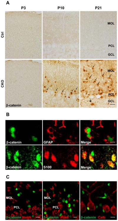
Progressive accumulation of beta-catenin in Bergmann glia during postnatal development. A. Representative images of the cerebellar cortex of control (Ctrl) and APC-CKO mice stained by beta-catenin. No accumulation of beta-catenin is identified in CKO mice at P3. Accumulation of beta-catenin becomes apparent but the vast majority of beta-catenin+ cells are located in the PCL at P10 (arrow), whereas many beta-catenin+ cells are distributed within the MOL at P21 (arrow). Note that a few beta-catenin+ cells are also identified in the GCL (arrowhead). B. Double immunofluorescence staining shows that beta-catenin accumulated cells are positive for astroglial markers GFAP and S100. C. None of beta-catenin+ cells is positive for a granule cell marker NeuN, an early granule cell progenitor marker Pax6, and a Purkinje cell marker calbindin (Calb). Scale bar: A, 20 μm, B, 5 μm, C, 10 μm. P, postnatal day; PCL, Purkinje cell layer; MOL, molecular layer; GCL, granule cell layer.