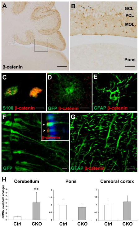
Figure 7.
APC deletion from astroglia induces beta-catenin accumulation and morphological changes in a region-specific manner. Representative images of sagittal sections of APC-CKO mouse brains at P21 stained by beta-catenin (A, B). B shows a magnified image of the boxed area in A. In contrast to apparent beta-catenin accumulation in the cerebellar cortex, no increase in beta-catenin immunoreactivity is observed in the pons. Note that beta-catenin+ cells are identified not only in the MOL/PCL but also in the GCL (arrow in B). Beta-catenin+ cells in the GCL are positive for an astroglial marker S100 (C). The reporter GFP+ and GFAP+ cells are broadly distributed in the pons of CKO reporter mice, but they do not accumulate beta-catenin (D, E). GFP+ tanycytes in the hypothalamus (F) and GFAP+ radial astrocytes in the white matter of the spinal cord (G) maintain their radial morphology and show no accumulation of beta-catenin in CKO reporter mice. Note that beta-catenin is apically concentrated (arrow head) in GFP+ tanycytes (inset in F) but is not accumulated within the nucleus (blue). Quantitative RT-PCR analysis shows that the expression of Axin-2 mRNA significantly increases in the cerebellum, but neither in the pons nor in the cerebral cortex of CKO mice at P21 compared to those of controls (H). **p<0.01, t test. n=4–6. Scale bar: A, 300 μm; B, 50 μm; C, D, E, 5 μm; F, G, 10 μm. P, postnatal day; PCL, Purkinje cell layer; MOL, molecular layer; GCL, granule cell layer.

