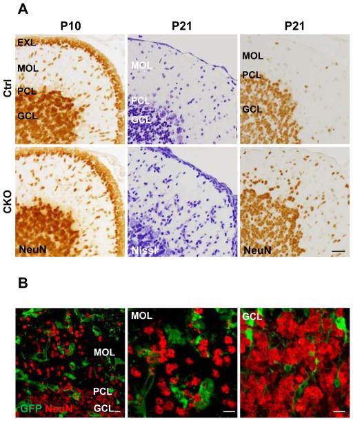
Figure 8.
Ectopic granule cells in the molecular layer of APC-CKO mice. A. Representative images of the cerebellar cortices of control (Ctrl) and CKO mice stained by Nissl and a granule cell marker NeuN. The thickness of the EXL and the number of migrating NeuN+ granule cells are indistinguishable between Ctrl and CKO mice at P10. The number of Nissl-stained cells in the MOL increases and a portion of them are positive for NeuN in CKO mice at P21, whereas only a few NeuN+ cells are identified in the MOL of Ctrl at the same age. B. Ectopic granule cells in the MOL are negative for GFP in CKO reporter mice at P21 (left and middle panels). Note that almost no GFP-positive granule cell is identified in the GCL as well (right panel). Scale bar: A, 25 μm; B, 10μm. P, postnatal day; PCL, Purkinje cell layer; MOL, molecular layer; GCL, granule cell layer; EXL, external granule cell layer.

