Figure 9.
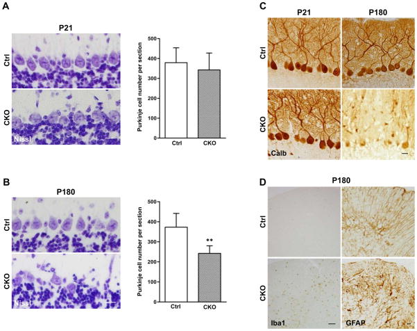
Cell non-autonomous neurodegeneration of Purkinje neurons and microglial activation in middle-aged APC-CKO mice. A, B. Representative images of the PCL of control (Ctrl) and CKO mice stained by Nissl and quantification of Purkinje cell number. At P21, the alignment of Purkinje cells in CKO mice is well preserved and the number is not significantly different (p=0.49). At P180, the loss of Purkinje cells becomes apparent and the number is significantly fewer in CKO mice. n=5, **p<0.01 versus controls, t test. C. Representative images of Purkinje neurons of Ctrl and CKO mice stained by calbindin. At P21, the dendritic arborization of Purkinje neurons in CKO mice is indistinguishable from that in Ctrl. At P180, in addition to a decrease in cell number, calbindin-immunoreactivity was markedly reduced in the remaining Purkinje cells. D. Representative images of the cerebellar cortices of Ctrl and CKO mice at P180 stained by Iba1 and GFAP. Iba1+ microglial cells are broadly distributed in the cerebellar cortex of middle-aged CKO mice. The distribution of GFAP+ astroglia remains unchanged in comparison to younger ages but GFAP-immunoreactivity moderately increases. Scale bar: : A, B, 15 μm; C, 20 μm; D, 50 μm. P, postnatal day; PCL, Purkinje cell layer; MOL, molecular layer; GCL, granule cell layer.

