
Figure 3.
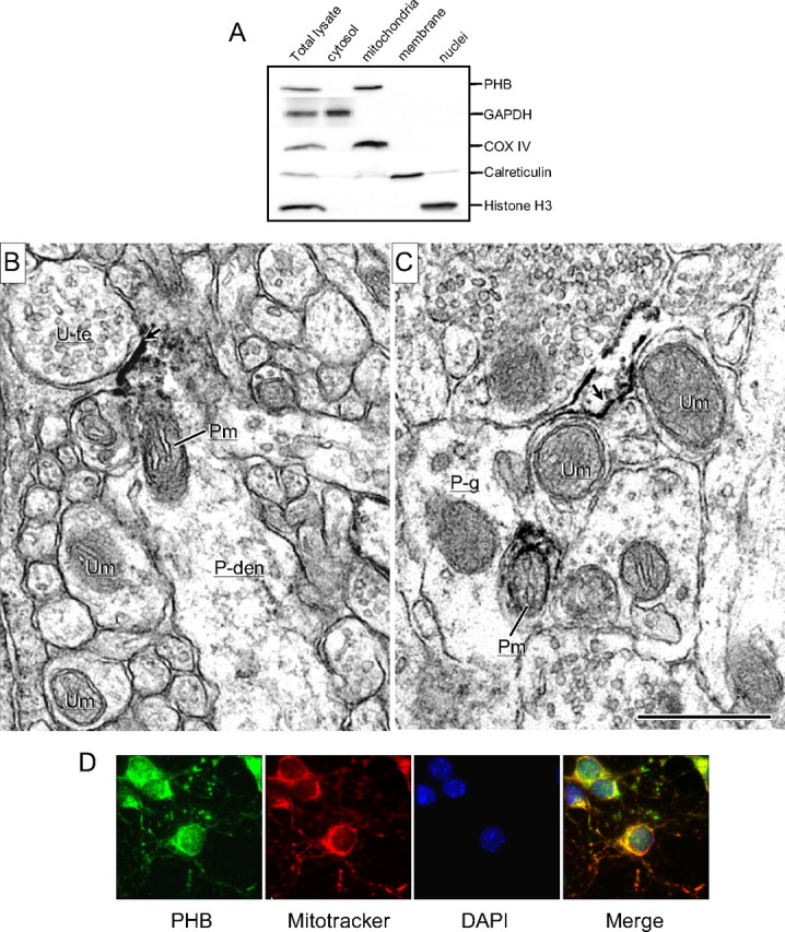
Neuronal localization of PHB. A, Subcellular fractionation of mouse neocortex. PHB is present mainly in the mitochondrial fraction and is not observed in the nuclear or cytosolic fraction. COX IV, histone H3, GAPDH, and calreticulin were used as mitochondrial, nuclear, cytosolic, and membrane markers, respectively. Results shown are representative from three separate experiments. B, C, Electron micrographs showing the immunoperoxidase localization of PHB in neuronal and glial profiles in the rat somatosensory cortex. B, PHB immunoreactivity (ir) is associated with inner and outer membranes of a mitochondrion (Pm) in a dendrite (P-den), located near an intensely labeled plasma membrane (arrow) contacted by an unlabeled terminal (U-te). Other neuronal processes within the neuropil contain unlabeled mitochondria (Um). C, PHB labeling is located on the plasmalemma (arrow) of a glial process (P-g) that sheaths small neuronal process containing mitochondria, which are either PHB-labeled (P-m) or unlabeled (U-m). Scale bar, 500 nm. D, In neuronal cultures, PHB immunoreactivity is colocalized with the mitochondrial marker MitoTracker Red.