Fig. 2.
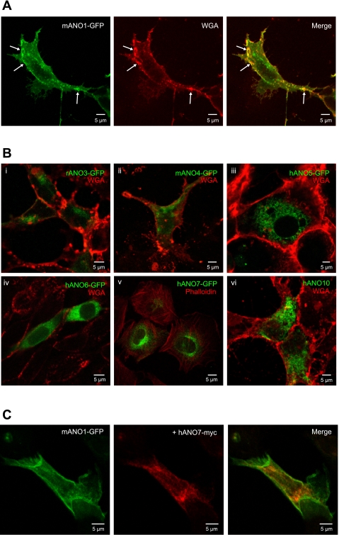
Immunofluorescence confocal microscopy of various cell types expressing ANOs 1, 3, 4, 5, 6, 7, and 10. A: HEK293 cells transiently transfected with green fluorescent protein (GFP)-tagged mANO1 alone. Arrows indicate colocalization of ANO1 with the membrane marker wheat germ agglutinin (WGA). B: various cell lines transiently transfected with GFP-tagged ANO constructs. Different cell types are shown for illustration, but these results are typical of all cell lines tested. i, ii, vi: HEK293 cells; iii, v: COS-7 cells; vi: CHO cells. Cells in B,v were counterstained with phalloidin for actin; all other cells were counterstained with a plasma membrane marker WGA. C: GFPtagged mANO1 (green) coexpressed with myc-tagged hANO7 (red) in HEK293 cell.

