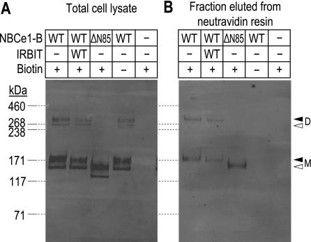
Fig. 4.
Comparison of the effects of amino terminal truncation and WT-IRBIT coexpression on NBCe1-B protein expression. A: representative Western blot of total oocyte lysates (samples are equivalent to 1/5th cell per lane) prepared from cells that were expressing full-length NBCe1-B (NBCe1-B/WT), full-length NBCe1-B + WT-IRBIT (NBCe1-B/WT + IRBIT/WT), truncated NBCe1-B (NBCe1-B/ΔN85), and cells that were injected with H2O following subjection to our biotinylation procedure (samples marked “+ biotin”). Samples marked “− biotin” were processed in the absence of biotinylation reagent. B: representative Western blot of biotinylated protein (equivalent to 1 cell per lane) that were isolated, using a neutravidin resin, from the same oocyte lysates that were sampled in A. Molecular mass markers are displayed to the left of A and are extended to B by dashed lines. The open triangles to the right of B indicate the migration position of the putatively non- or core-glycosylated BWT monomer (M) and dimer (D) visible in A, but not in B. The solid triangles indicate the migration position of putatively complex-glycosylated BWT M and D visible in both A and B.

