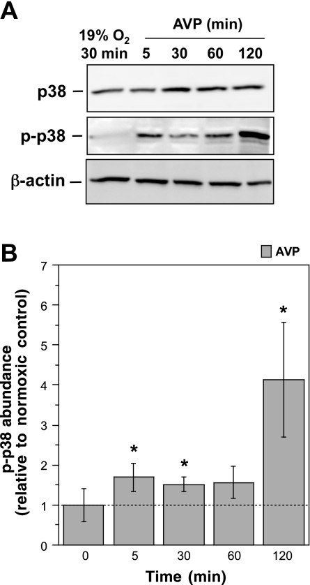
Fig. 3.
AVP-induced activation of p38. CMEC monolayers were exposed to control normoxic medium (glucose-containing HEPES-DMEM) or AVP (100 nM) in normoxic medium for 5, 30, 60, or 120 min. Cell lysates were subjected to Western blot analysis using antibodies that recognize p38 (total kinase) or p-p38 (phosphorylated, active kinase). A: representative Western blots. B: p-p38 abundance. Values are means ± SE of 7 separate experiments. *Significantly different from control, P < 0.05 by 1-tailed paired t-test.

