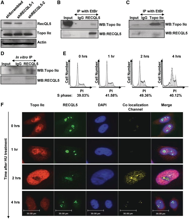
Figure 4.
RECQL5 physically interacts with Topoisomerase IIα during S-phase. (A) Western blot of whole cell lysates from HeLa shScrambled, shRECQL5-1 and shRECQL5-2 cells, probed with indicated antibodies. Equal loading was confirmed by probing with anti-Actin antibody. (B) Immunoprecipitation of RECQL5 and (C) Topoisomerase IIα from HeLa whole cell extracts and probed with indicated antibodies. (D) Immunoprecipitation of RECQL5 from a mixture of RECQL5 and Topoisomerase IIα recombinant proteins and probed with indicated antibodies. (E) Representative flow cytometry histograms of HeLa cells analyzed after 2 mM hydroxyurea treatment and released for the time indicated. DNA content stained with PI was plotted against cell number. (F) Confocal images of representative HeLa cells fixed and stained for RECQL5 and Topoisomerase IIα after 2 mM hydroxyurea treatment and then released for the time indicated. Staining protocol is described in ‘Materials and Methods’ section. Images are pseudo colored: green-RECQL5, red-Topoisomerase IIα and blue-DAPI stain of nucleus, with yellow areas indicating the co-localization. The co-localization channel was generated by the Volocity software. Co-localization was determined by the following equation: (Xi −Xmean) (Yi −Ymean) where Xi is the intensity of the voxel for the Red Fluorescence Channel and Yi is the intensity of the voxel for the Green Fluorescence channel (62). The fifth panel is the merge of all three channels.

