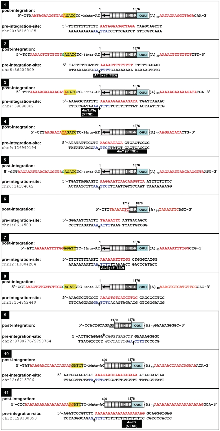
Figure 5.
Genomic SVAE de novo insertions display hallmarks of L1-mediated retrotransposition. Both pre- and post-integration sites are presented. Insertions 1–8 and 9–11 are derived from SVAE reporter elements encoded by pAD3/SVAE and pAD4/SVAE, respectively. Nucleotide positions at the 5′-end of each SVA insertion refer to the reference sequence of SVA H19_27 [(21); Supplementary Figure S1]. Marked full-length insertions cover ~3.3 kb (pAD3/SVAE-derived) and ~2.8 kb (pAD4/SVAE-derived), respectively. Identified 5′-truncated insertions comprise 1580 bp (insertion 6) and 1620 bp (insertion 9), respectively. The L1EN cleavage site on the bottom strand is indicated in blue. Extra deoxyguanylates at the 5′-ends of de novo insertions are indicated in green. CMVP-derived sequences are highlighted in yellow. Nucleotides representing patches of microcomplementarity are underlined. Black bars, Alu TSD sequences. Red lettering, SVA TSD sequences; neo, neomycin-phosphotransferase gene.

